Features
If you are new to petrographic microscopy, start here!
This section introduces some key properties of minerals that can be used to help identify and understand them in thin section. If you are unfamiliar with rocks under the petrographic microscope, this is a good place to start discovering how to observe and interpret what you see in the microscope images. As you gain confidence in recognising these aspects of minerals, you might like to move on to exploring minerals and textures in the other sections linked on the right of this page. If you find something weird, you could see if it is one of the artefacts or defects we've called 'Tricksters' that have puzzled all microscopy students at some stage!
How to use this page
Click on each coloured bar below to expand details on that item, including a brief explanation of the property and a button linking to an example view of a Virtual Microscope sample. There is also a screenshot of the relevant feature below the button for a quick view. If the screenshot is square, the link button will take you to a microscopic view of the thin section, zoomed in to the feature described. Circular screenshots indicate that the link button will take you to a rotation view; many of the features listed here require rotation of the stage to be observed.
Features seen in plane polarised light (PPL) are listed first, followed by those only seen under crossed polars (XPL).
PLANE POLARISED LIGHT (PPL)
This is the standard lighting mode for a petrographic microscope, with a polarising filter between the light source and the thin section of rock. The interaction of the polarised light rays with the different minerals in the thin section produces optical effects that can aid in mineral identification. It also allows observation of the rock textures and microstructures caused by deformation, mineral reactions and other processes in the Earth's crust.
- RELIEF
Relief describes how sharply a mineral stands out in thin section compared to the background. In this view, the pinkish-grey mineral (garnet) has the highest relief, followed by the bluish green/yellow mineral (amphibole), the brown mineral (biotite) and finally the colourless groundmass (plagioclase). There are also small, roundish colourless grains of apatite that have higher relief than the plagioclase.
Tip: High relief minerals typically appear cloudy and greyish, rather than clear.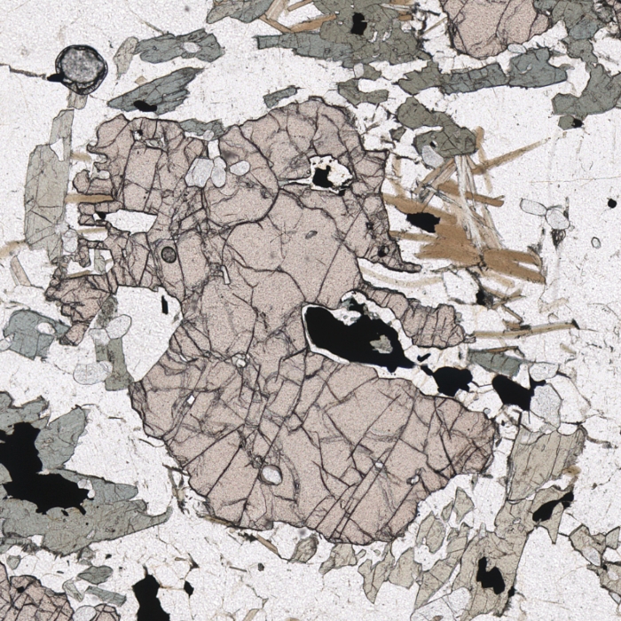
- RELIEF - Twinkling
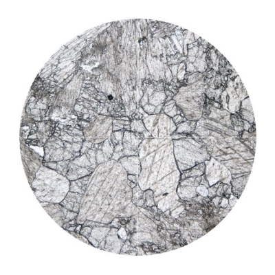
A few minerals show contrasting relief in different orientations, because their refractive index changes with orientation. This is perhaps most marked among common minerals in calcite. The example here shows a cluster of colourless calcite grains. As you rotate the image through 90°, note how the calcite grains change appearance in the plane polarised view: clear, white grains become cloudy grey (higher relief), while greyish grains become clear (lower relief).
- CLEAVAGE - Prismatic
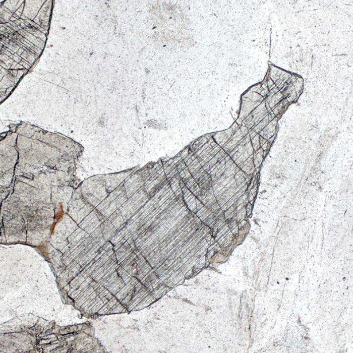
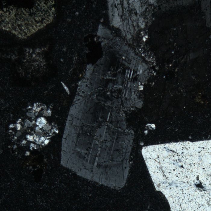
Cleavage appears as a pattern of lines on prismatic crystals of minerals such as mica, amphibole and pyroxene. Cleavage that develops parallel to the length of the prism is called prismatic. A prismatic cleavage means that during rock cutting and thin section manufacturing, the prismatic mineral was cut parallel to the prism development (long, crystallographic c-axis). The cleavage patterns are forming during crystallographic growth when repetitive planes of atoms or ions define detachment surfaces on a crystal.
The rock linked here has numerous examples of pyroxene crystals with prismatic cleavage. Contrast these with olivine crystals in the same sample, which have no cleavage but instead are cross-cut by irregular, dark cracks.
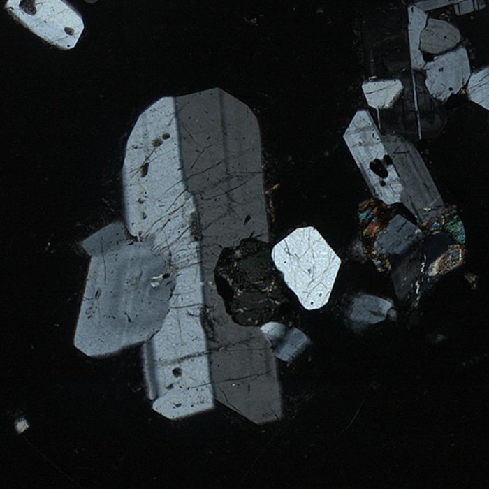
- CLEAVAGE - Intersecting sets
Intersecting sets of cleavage in prismatic minerals are apparent when a crystal is cut perpendicular to its prismatic growth, so that the traces of 2 or more different prismatic cleavages are intersecting on the mineral surface. In amphibole there are 2 parallel cleavages that intersect at 120° angle, and in pyroxene, as in the example below, it is nearly orthogonal (90° angle). Other minerals where intersecting sets of cleavage can be seen are calcite and kyanite.
- PLEOCHROISM
Pleochroism describes the tendency of some minerals to change colour under plane polarised light as the stage is rotated. Many change colour from a dark shade of one colour to colourless, but some may show three colours for grains in particular orientations.
Perhaps the commonest mineral to display strong pleochroism is biotite. Most commonly, biotite shows pleochroism from rich brown to colourless – though this depends on the orientation of the crystal. This rotating view shows several biotite grains in different orientations. The more prismatic (elongate) the grain appears, the more extreme its pleochroism, from dark brown to almost colourless. Less elongate grains show less change, from dark to pale brown, while one grain on the edge of the view barely changes colour. When viewed exactly down the crystallographic optic axis of the mineral, there will be no change in the brown colour (and under crossed polars, the grain will be in permanent extinction.)
- PLEOCHROISM - Three colours
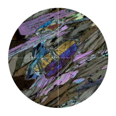
Some minerals display pleochroism in three colours, corresponding to the three different crystallographic axes. This example shows several crystals of the amphibole glaucophane in different orientations, each showing distinct colour changes: blue to lilac (long, prismatic crystal); blue to colourless (elongate grains at top and lower right of view); lilac to colourless (stubby grains clustered round centre of view).
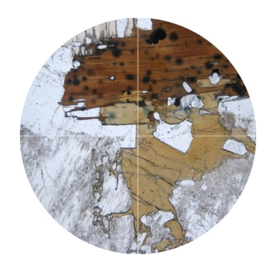
- PLEOCHROISM - Pleochroic haloes
In the pale brown biotite in the top left quadrant of this rotating view, there are a few small inclusions of opaque and colourless minerals – but also some fuzzy grey blobs. As you rotate the view and the biotite darkens, these grey blobs also darken and become more distinct, appearing as black circles with fuzzy edges. Known as ‘pleochroic haloes’ these features occur in a few minerals, indicating radiation damage from tiny inclusions of U- or Th-bearing minerals like zircon or monazite. In some cases, the inclusion is visible, but for many the only clue to their presence is the circular pleochroic halo in the host mineral.
CROSSED POLARS (XPL)
In this lighting mode, an additional polarising filter is slid in at right angles to the first polariser. This reduces the amount of light reaching the observer, so microscopic views in XPL tend to look much darker than PPL views. However, minerals under crossed polars display many features not visible in PPL mode, aiding identification and revealing hidden aspects of their crystal structure and chemical composition.
- EXTINCTION - Parallel
Extinction occurs in most minerals under crossed polars when the mineral turns completely black as the stage is rotated. In most orientations, a crystal will go into extinction four times in a single rotation of the stage (every 90°). If the crystal is oriented to be viewed directly down the c-axis, it will appear permanently black on rotation (like an opaque mineral).
The long axis of this kyanite crystal under the cross hairs is initially at 45° to the polarizer (usually mounted E-W). Rotating 45° clockwise or anticlockwise brings it to an extinction position (black under crossed polars). At this point, both the cleavage and the straight edges of the crystal are aligned either N-S or E-W, so it is said to have straight, or parallel, extinction.
- EXTINCTION - Inclined
Minerals that go into extinction when their cleavage (and hence straight crystal edges) are not orthogonal (E-W or N-S) but at an angle are said to show inclined extinction. The plagioclase grains in this rotation view are good examples. In all cases, they turn black when inclined at quite a high angle to the orthogonal directions. Many of these crystals have simple twins, and both halves of the crystal show inclined extinction.
- EXTINCTION - Birdseye
A few minerals do not turn completely black in extinction, but instead have a mottled black appearance, rather like a starry sky. The example shown is a grain of calcite that is almost in extinction (very dark but not perfectly black). The bright speckles across the grain are not tiny inclusions; they are typical of birdseye extinction. This type of extinction is also a feature of micas (biotite, muscovite).
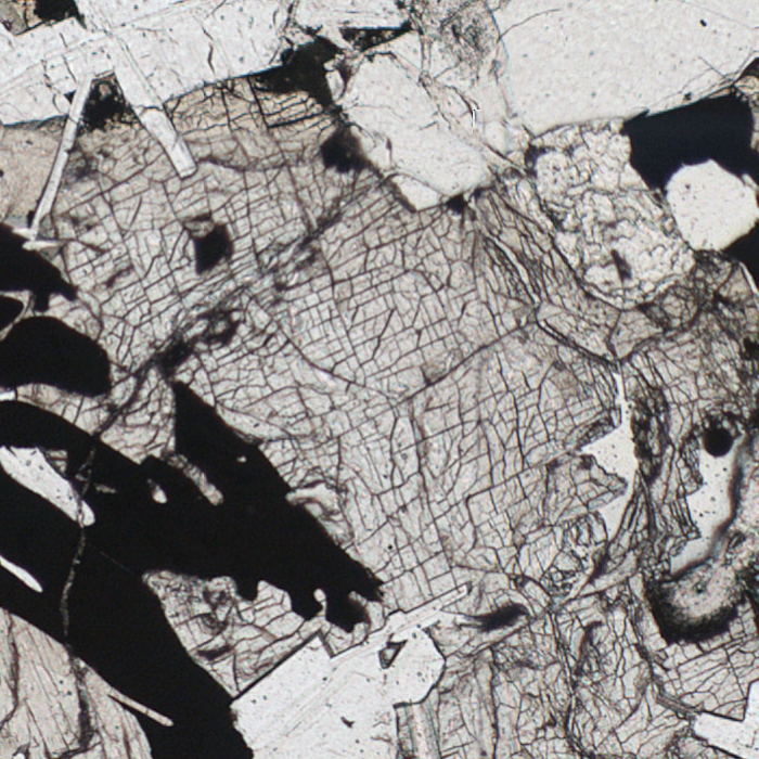
- EXTINCTION - Undulose
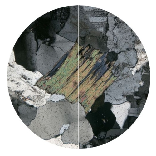
Undulose extinction is a special state of extinction when the minerals have been subjected to dynamic deformation. Such deformation can damage the crystal structure of minerals, creating defects in the lattice. In this case, this damage is revealed by undulose extinction, where a mineral grain does not turn completely extinct (i.e., black) all at the same time: irregular patches show different shades of grey. A common mineral that presents undulose extinction is quartz, but other affected minerals can be pyroxene, amphibole or micas. In the displayed example, there are quartz grains displaying undulose extinction to the upper left and lower left of the central mica crystal. Other examples are visible throughout the thin section in the static view.
- TWINNING - Simple
Sometimes single crystals under crossed polars are divided in two. This phenomenon in petrography is called twinning and results in two different "areas" in a single crystal that share different extinction angles and usually different birefringent colours (e.g., in pyroxene or in amphibole). In fact, twin crystals results from the intergrowth of two adjacent crystals whose crystal lattice have a common point. Twin crystals are characterized by symmetrical patterns.
- TWINNING - Lamellar
Lamellar twinning occurs when there are multiple "simple" twin crystals in a repeating pattern in a mineral. The results in crossed polars is a single crystal but with multiple parallel "stripes" that each one extinct in a different angle. The mineral that always exhibits lamellar twinning under microscope is plagioclase, making it very easy to recognize in a thin section.
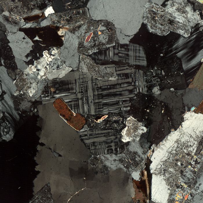
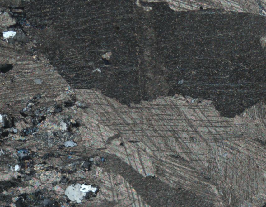
- TWINNING - Microcline (cross-hatched)
Microcline twinning (i.e., cross-hatched twinning) is a specific type of twinning occurring in alkali-feldspar and especially in microcline. Microcline is the variety of alkali-feldspars, occurring in plutonic igneous rocks that had sufficient time for crystallization resulting in well-structured lattices. Microcline twinning under crossed polars exhibits twin lamellae which crosscut each other in a crossing pattern.
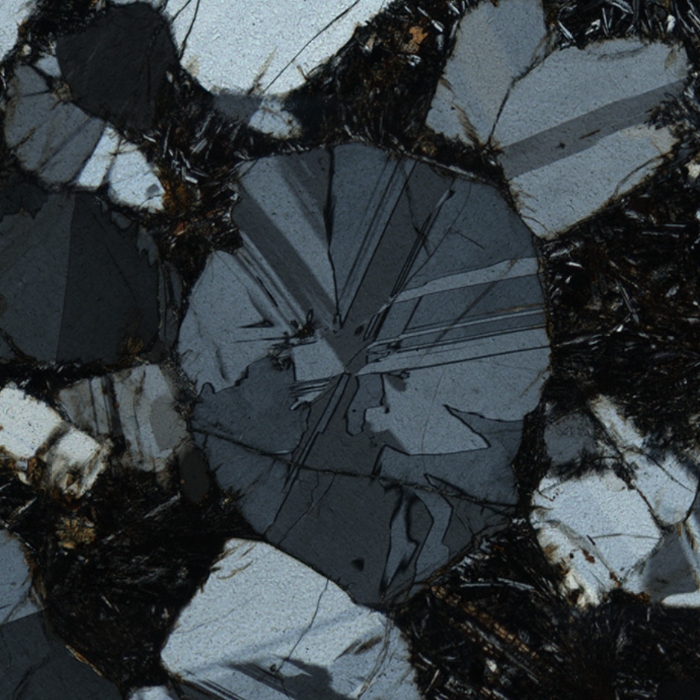
- TWINNING - Cyclic
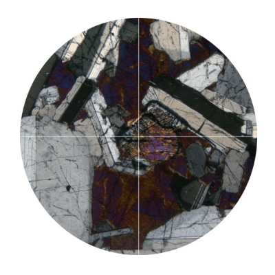
In cyclic twinning, segments of colour appear to radiate from a common centre and fan out radially, like a pizza cut into slices. In this view under crossed polars, a roundish cordierite crystal displays cyclic twinning of this form. Adjacent twins are different orientations and so appear in different tones of grey, forming the characteristic pattern.
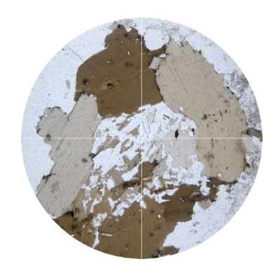
- ZONING - Monotonic
Zoning patterns are observed in magmatic or metamorphic rocks when there are differentiations in the composition of a magma or a metamorphic rock during crystal growth. The typical zoning is defined by a mineral which has different composition at its core than its rim. The different composition in the mineral's core and rim, are representative of the formation conditions at that time. Typical monotonic zoning is observed in minerals with many endmembers such as, garnet, pyroxene, amphibole and plagioclase. Below, a plagioclase crystal under crossed polars where at it's rim the birefringence changes as a consequence of a change to mineral composition.
- ZONING - Oscillatory
Some zoning occurs as fine, alternating bands of different composition, visible as different interference colours under crossed polars. This oscillatory zoning reflects rapid, fluctuating changes in conditions as the crystal grows, more typical of crystals growing from magma. This example shows oscillatory zoning in a plagioclase crystal, displayed as concentric, alternating bands of light and dark grey.

- INTERFERENCE COLOURS - Low
In petrography and optical microscopy, petrographers use a colour chart in order to better distinguish and recognize the minerals that make up a rock. The most famous one is called Michel-Levy chart, which was developed by Auguste Michel-Levy. Given the thickness of a thin section (usually this is set at 30 micrometres), the interference colours are divided in three orders depending on their intensity. The interference colours are visible due to an optical effect of the crystals, when the cross-polarized light come through them, called birefringence. Birefringence is measured by the difference in speed between the fast and slow light rays.
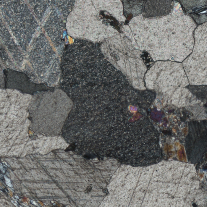
Minerals with low interference colours show a variety of greyish hues as well as first order yellow and red colours. These minerals are quartz, feldspar, plagioclase, orthopyroxene, apatite, cordierite and others. Below is an assemblage of quartz grains with low interference colours from a granite.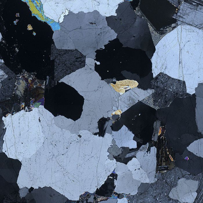
- INTERFERENCE COLOURS - Moderate
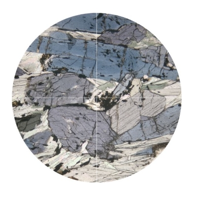
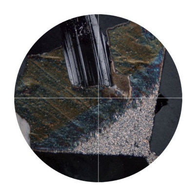
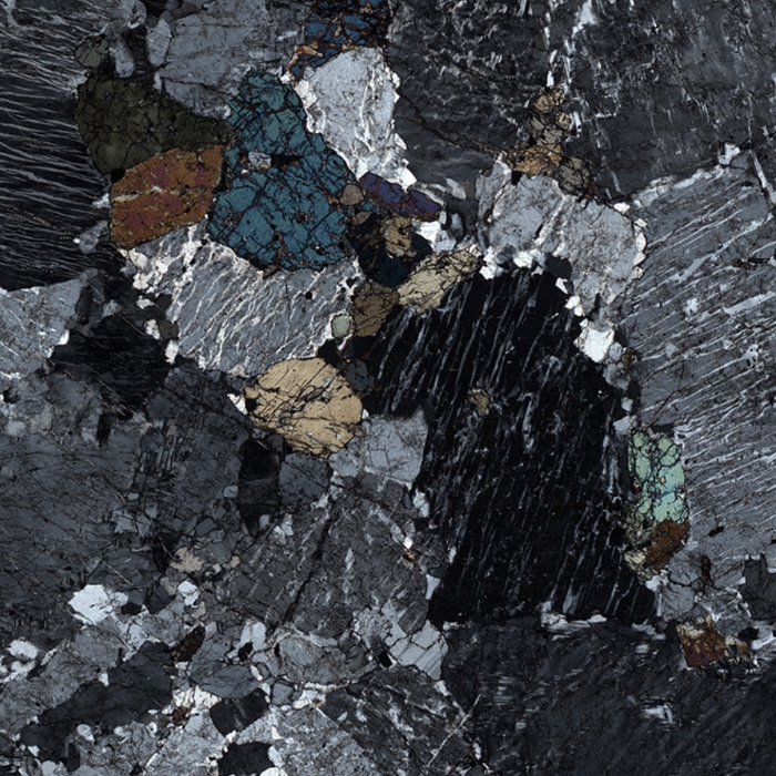
Moderate interference colours do not contain greyish colours in their range; instead they range through the 'first order' rainbow spectrum and into the lower part of the second order, including yellow, brown, red, magenta and blue. Typical minerals that have moderate interference colours are hornblende and kyanite. Some other amphiboles and clinopyroxenes may exhibit such colours (depending on the cut of the crystal).
The image below shows grains of amphibole and pyroxene with a range of moderate interference colours from creamy grey to second-order blue.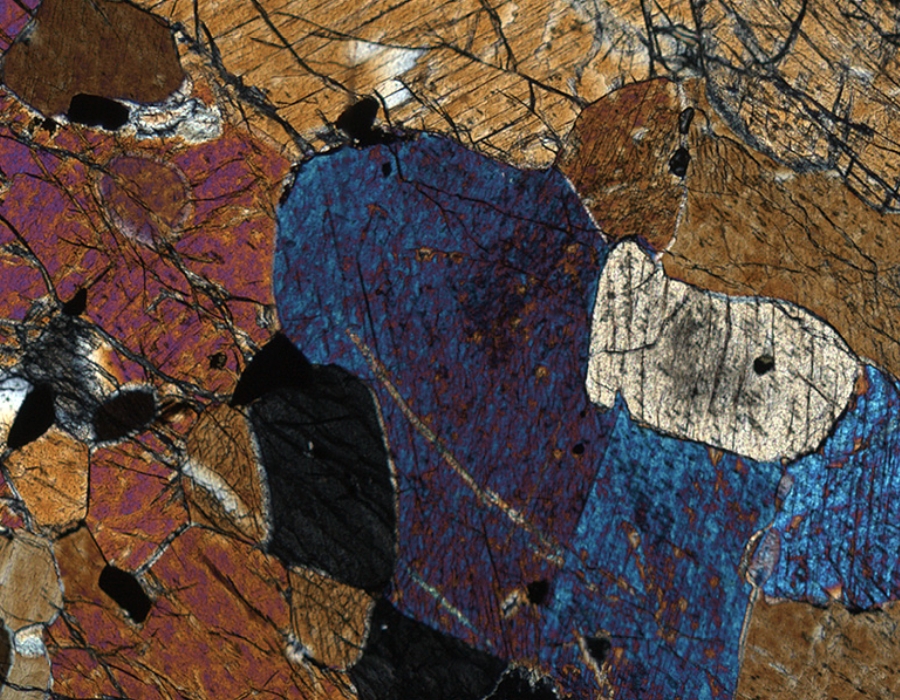
- INTERFERENCE COLOURS - High
High interference colours lie further up the Michel-Levy scale than moderate interference colours. The former are vibrant, vivid, intense colours that appear very bright on the mineral. Minerals that may show high interference colours include olivine, biotite mica, muscovite mica, and epidote.
The example below (and linked above) shows a shocked olivine from a lunar sample, with interference colours running up to third order lime green. The series of rainbow stripes just right of centre occur on a sloping edge of a crystal: the thickness variation from almost zero to the standard 30 microns results in bands of interference colour from first order grey to second order pink.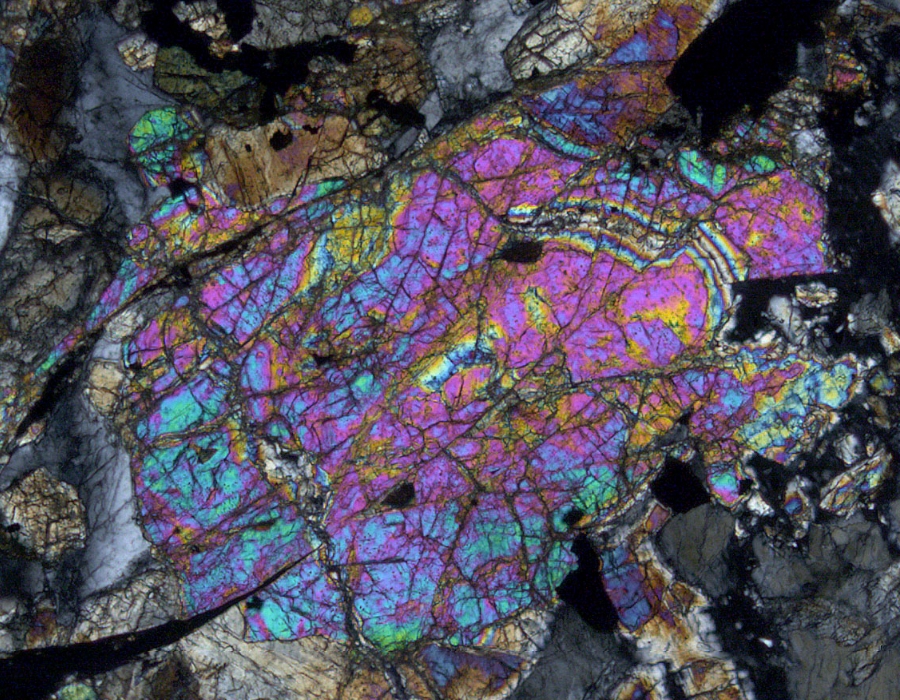
- INTERFERENCE COLOURS - Very High
Very high interference colours are typically extremely iridescent, marking an even more amplified case of the effect noted for high interference colours. Very high interference tends to be so bright that specific colours become harder to separate and the crystal begins to look like it has 1st-order (low) interference (white, greys, black) covered with an iridescent effect. The most common example of this effect is seen in the mineral calcite (below), but it can also be encountered in titanite (sphene). Titanite shows a slight variation on this effect when the high interference overwhelms itself and the crystal shows the same colour that it did in plane-polarised light.
- EXSOLUTION - Perthite
In this example, under crossed polars, several alkali feldspar grains of different shades of grey show irregular streaks of paler grey across the crystal. This is a typical perthite texture: the irregular streaks are sodic plagioclase feldspar that has exsolved from the alkali feldspar during cooling.
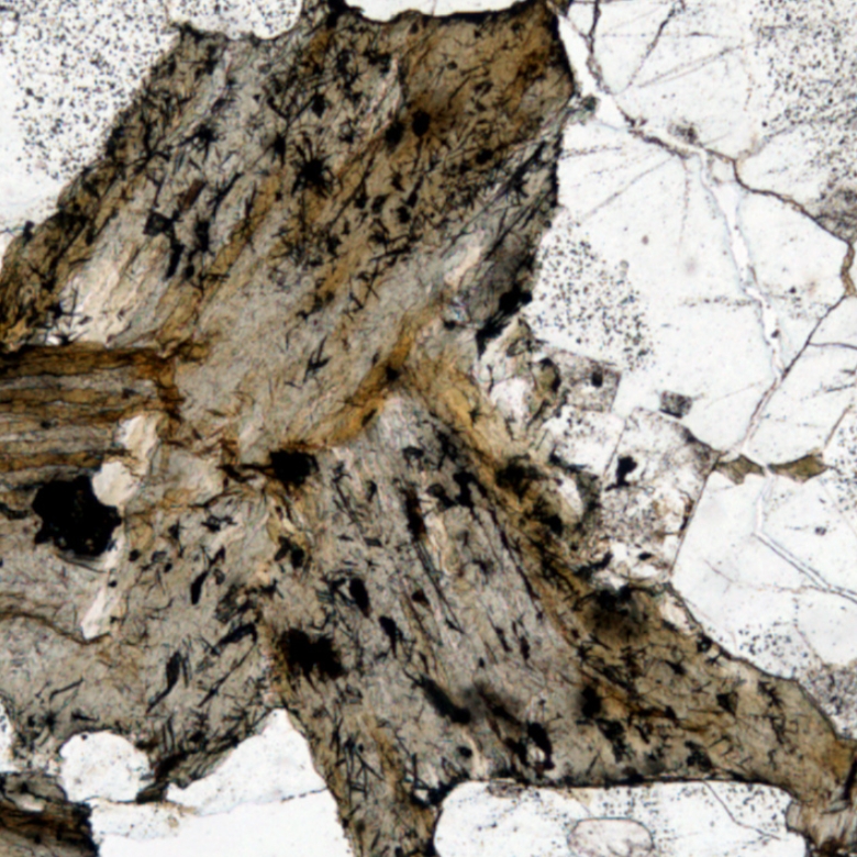
- EXSOLUTION - Rutile in biotite
Some biotite grains are very rich in titanium - especially those formed at high temperatures. Titanium ions are much more compatible in mafic minerals than in felsic minerals and melts. This means that, in felsic melts, when the more mafic phases, such as biotite, crystallise, the titanium will tend to concentrate in those minerals. However, titanium is incompatible in chlorite. This means that, when biotite alters to chlorite, the titanium from the biotite cannot be accommodated in the chlorite lattice and has to exsolve, forming a different phase. This is typically rutile, or titanium oxide (TiO2), which forms dark brown needles within the altered biotite. In some cases, the rutile needles form at 60° to each other, their orientation apparently controlled by the crystal lattice of the biotite. There is some suggestion of this pattern in some of the rutile needles in the image below.