Tricksters
If you've found something weird in a thin section that you cannot identify, it's worth checking out these oddities.
CAUTION: work in progress! Please be patient while the VM elves scurry through our Collections looking for good examples of these gremlins to fill in the gaps.
If you do find a good example while browsing our VM Collections, please email virtual-microscope@open.ac.uk and let us know. Contributions to this page will be duly attributed!
- ABRASIVE PARTICLES
To grind down and then polish thin sections of rock, abrasive powders (usually in a fluid slurry) are used. Sometimes, these particles can get trapped on the surface and are not removed when it is cleaned.
In this example, many quartz grains in the sample are indurated with carborundum particles, giving them an unusual, speckled appearance in plane polarised light. Some grains show clear rims. Other minerals (feldspar, white mica, biotite) appear unaffected.
These could be mistaken for inclusions of fluid or other minerals in the quartz, but one clue is that the particles have a very consistent size (due to the grade of the abrasive powder) – inclusions would vary in size, shape and colour. Dusty alteration of the feldspar grains also looks different: finer-grained, patchy, and with interference colours visible under crossed polars.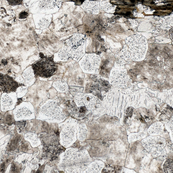
- ABRASIVE PARTICLES - Patches
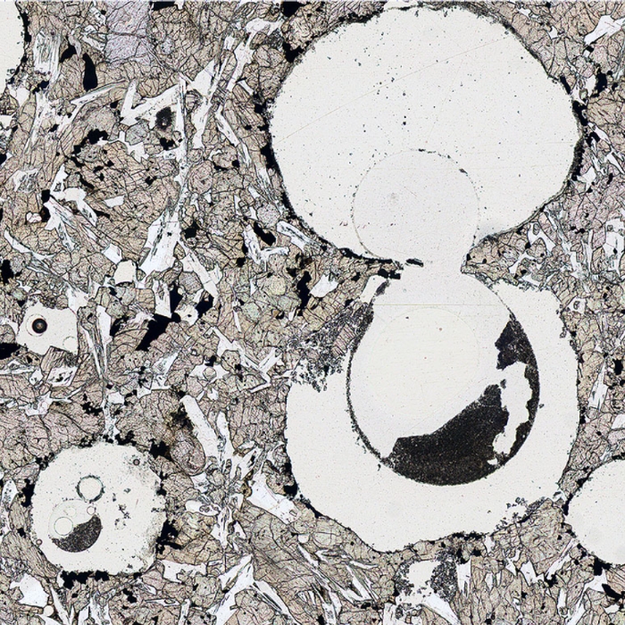
In some thin sections, patches of abrasive powder are trapped in cracks or voids. These are visible as irregular dark masses of material. This example shows a large and a small patch of abrasive trapped in large air bubbles that were themselves trapped when this sample was impregnated with resin. This impregnation was done because the basalt is vesicular – full of gas bubbles – and hence liable to break up during grinding and polishing.
- AIR BUBBLES
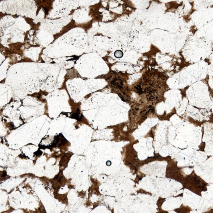
Occasionally, as the rock chip is glued to the glass slide, tiny amounts of air are trapped as bubbles. These appear as dark-rimmed features, typically clear inside and commonly (but not always) circular. The view here shows two bubbles in a thin section of a sandstone.
- CRACKS IN GLASS SLIDE
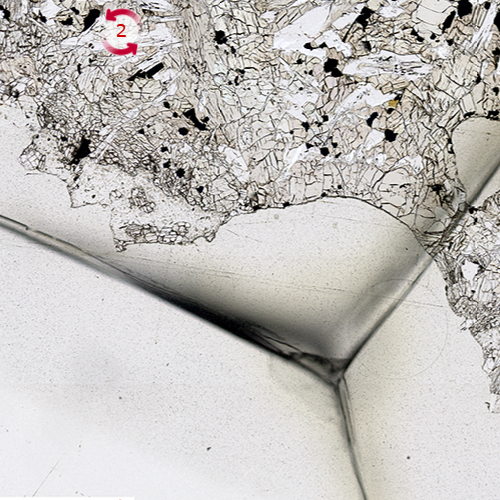
Accidents happen, and unlike virtual microscope slides, real slides are made of glass so they break completely or the coverslip breaks (they are very thin). Generally broken slides are thrown away but some slides are too precious to throw away there are rare cracked slides in our collections. Most of the cracks you'll find are in the Apollo Moon rocks because...well... no one has been there since 1972 so we can't just get a spare. This is a good example where the cover slip has broken but everything else is ok so we scanned it anyway.
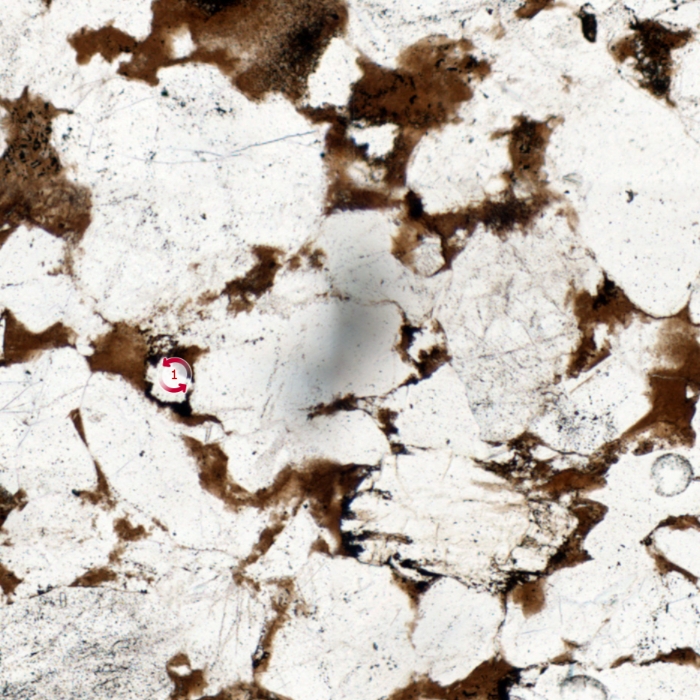
- DIRT
If slides are not carefully cleaned before viewing, various substances on the slide can appear as dark, indistinct smudges. This example is blurred, suggesting it is a tiny patch of dirt on the lower surface of the glass slide which is out of focus. You can also view this feature on the rotation view labelled (1); it does not change appearance on rotation.
- DUST PARTICLES
Many thin sections - and some rocks - contain 'holes': areas where there is no rock glued to the glass slide. These appear very dark or black under crossed polars, but sometimes you may spot a dark circle with a dark cross within, as in the view below. This generally marks a speck of dust or minute rock particle, caught in the resin that filled the hole. These artefacts are a good clue that the dark patch is not a strangely-shaped garnet after all, but a ragged hole in the sample. These may be natural (vugs, vesicles or pores), or might be formed during thin section polishing when a crystal is plucked out of the surface of the slide.
A bonus feature in the image below is the series of rainbow stripes on the edge of the vivid olivine crystal below the dust particle, which marks the bevelled edge of the crystal. These show how the interference colours depend on the thickness of the mineral, running from first order grey (upper left, thinnest) to the maximum bright green (lower right of crystal, thickest).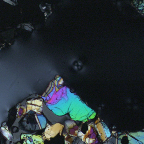
- FIBRES
The VM elves are hunting for a good example of this trickster. Please check back in a while!
Alternatively, if you find a good example in one of the VM Collections, please email us at virtual-microscope@open.ac.uk and let us know. Contributions to this page will be duly attributed!
- HOLES, VOIDS
The VM elves are hunting for a good example of this trickster. Please check back in a while!
Alternatively, if you find a good example in one of the VM Collections, please email us at virtual-microscope@open.ac.uk and let us know. Contributions to this page will be duly attributed!
- PEN MARKS
Occasionally, as the rock chip is glued to the glass slide...
- SCRATCHES
The VM elves are hunting for a good example of this trickster. Please check back in a while!
Alternatively, if you find a good example in one of the VM Collections, please email us at virtual-microscope@open.ac.uk and let us know. Contributions to this page will be duly attributed!
- THICKNESS VARIATION
The VM elves are hunting for a good example of this trickster. Please check back in a while!
Alternatively, if you find a good example in one of the VM Collections, please email us at virtual-microscope@open.ac.uk and let us know. Contributions to this page will be duly attributed!